2026-09-04
Contenido
Put a prepared onion-skin slide under a school light microscope and, at 400× magnification, students can trace cell walls and find the darker nuclei inside each cell. Put the same tissue under a transmission electron microscope and the field of view opens up to mitochondria, ribosomes, and the double membrane around the nucleus. That jump in visible detail is the shortest possible answer to this question: an optical microscope builds its image from visible light focused by ground glass lenses, while an electron microscope builds its image from a beam of electrons steered by electromagnetic lenses. Every other difference — resolution, magnification, specimen preparation, size, running cost, even the color of the final image — follows from that one substitution.
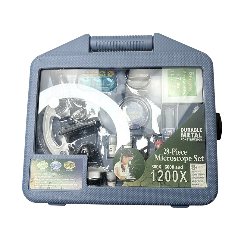 Biology Student Microscope Kit with Slides and Tools Offering 300x–1200x magnification with metal, plastic, and glass construction, this kit includes blank and prepared slides, a magnifier, and dissection tools, letting students perform the hands-on wet-mount and slice observations the passage describes for light microscopy. View Product →
 Resolution — the ability to show two nearby points as separate — is limited by the wavelength of whatever beam does the imaging. Visible light spans roughly 400 to 700 nanometers, and even a perfectly built optical system cannot resolve detail much finer than about half of that. In practice, the ceiling for a well-aligned compound light microscope sits near 200 nanometers.
Electrons behave as waves too, but their wavelength is vastly shorter. An electron accelerated through 100,000 volts has a wavelength of about 0.0037 nanometers — more than 100,000 times shorter than green light. As the chart shows, the wavelength keeps shrinking as accelerating voltage rises, which is why high-voltage instruments can resolve individual atomic columns.
Because glass cannot focus charged particles, an electron microscope replaces lenses with electromagnetic coils, and the entire beam path runs inside a high vacuum — stray air molecules would scatter the electrons long before they reached the specimen. A light microscope faces no such constraint and works happily on an open bench.
The table pulls the main specifications together; each row is expanded in the sections that follow.
| Feature | Optical (Light) Microscope | Electron Microscope |
|---|---|---|
| Imaging beam | Visible light, 400–700 nm | Electron beam, about 0.004 nm at 100 kV |
| Focusing method | Ground glass lenses | Electromagnetic coils |
| Maximum useful magnification | 1,000–1,500× | 100,000× (SEM) to over 1,000,000× (TEM) |
| Best resolution | About 200 nm | About 1 nm (SEM); 0.1–0.2 nm (TEM) |
| Specimen condition | Live or fixed; wet mounts welcome | Fixed, dried, coated or resin-embedded; never alive |
| Environment | Open bench, normal air | High vacuum, dedicated room |
| Image color | Natural color | Grayscale; false color sometimes added |
| Operator training | Minutes to hours | Weeks to months |
On paper, the numbers look lopsided. A good student compound microscope tops out around 1,000 to 1,500× before the image simply grows blurrier — a phenomenon called empty magnification, because pushing past the 200-nanometer resolution limit makes everything bigger but reveals nothing new. A scanning electron microscope (SEM) routinely reaches about 100,000×, and a transmission electron microscope (TEM) can pass one million times.
Read the resolution chart with its units in mind: at 0.2 nm, a TEM distinguishes features about a thousand times finer than a light microscope. That is the gap between seeing a cell nucleus and seeing ribosomes dotted along its outer membrane. For scale, a human hair is roughly 80,000 nm across — easy work for a light microscope. A virus at 20–300 nm sits right at the boundary, and a DNA double helix at 2 nm will never appear in an optical image.
This is the difference most people underestimate. A light microscope accepts a wet mount made in under a minute: a drop of pond water, a cover glass, done — and the organisms keep swimming. Simple stains such as iodine or methylene blue add a few minutes and make nuclei and starch grains stand out. Students can prepare their own slides, which is half the pedagogical value of the exercise.
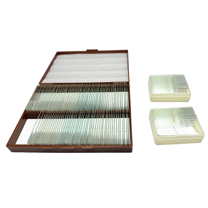 Biology Education Slide Set for Cell and Tissue Study This glass and plastic kit provides botany, zoology, anatomy, and general biology prepared slices for studying cells and tissues at the microscopic level, complementing the discussion of staining and specimen preparation with ready-made samples. View Product →
 An electron microscope imposes the opposite regime. The vacuum rules out anything wet or living: specimens are chemically fixed and dehydrated first. For SEM imaging they are then mounted and sputter-coated with a whisper-thin film of gold or palladium to carry the beam charge away. For TEM they are embedded in resin, sliced into sections just 50–100 nanometers thick on an ultramicrotome, and stained with heavy metals. Preparation routinely takes hours, demands trained hands, and can introduce artifacts — details that show up in the image but were never part of the living cell.
For schools, the curriculum decides. Secondary biology syllabi ask students to observe onion and cheek cells, leaf cross-sections, mitosis in root tips, and pond microorganisms — all comfortably within a 40× to 400× range, with 1,000× oil immersion reserved for bacterial smears in advanced classes. An optical microscope delivers every one of those observations, teaches core lab-handling skills, and survives daily use by teenagers.
Electron microscopy, by contrast, belongs to universities, research institutes, and industrial labs. What schools do need is conceptual understanding: exam questions routinely ask why electron microscopes revealed organelles that light microscopes could never show, and how the two instruments compare. When the syllabus reaches that unit, a purpose-built teaching microscope makes the lesson concrete.
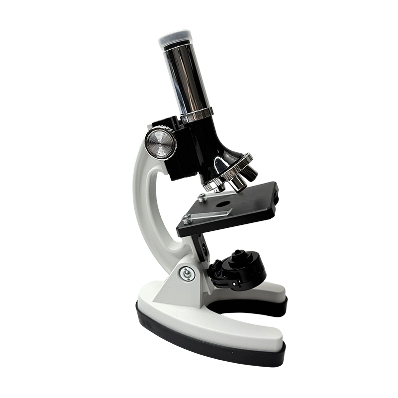 School Laboratory Biological Microscope for Teaching With magnification options from 200X to 1000X and standard components like objective converters and coarse and fine focusing, this precision optical microscope suits observing cells, bacteria, and slices, making classroom lessons comparing light and electron microscopy concrete. View Product →
 When equipping a laboratory, it pays to plan from the full range of biology teaching instruments rather than the microscope alone — prepared slides, anatomical models, and dissection kits turn a magnifier into a complete course.
No. The vacuum and the electron beam itself destroy living tissue, and specimen preparation kills the sample before it ever enters the column. Living processes are observed with light-microscopy techniques such as phase contrast.
Electrons carry no color information. Detectors record signal intensity — how many electrons arrive, and from where — so raw images are grayscale. The vivid colors in published micrographs are added afterward to label different structures.
No. A stereo, or dissecting, microscope is still an optical instrument: it uses visible light to give a lower-magnification, three-dimensional view of whole objects such as insects or plant parts. It is often the first microscope younger students meet.
A 40×, 100×, 400× turret, with 1,000× oil immersion available for advanced work, covers most secondary biology. Beyond that, extra magnification on a light microscope adds no detail — the 200 nm resolution limit, not the eyepiece, sets what can be seen.
The two microscopes are not rivals so much as complements aimed at different scales: one renders the living, colored world of the cell; the other maps the molecular machinery inside it. For procurement teams, the practical rule is straightforward — buy optical microscopes to match what the syllabus asks students to see, and treat electron microscopy as a concept to teach and a facility to visit. For ideas on structuring hands-on lessons around existing equipment, this guide on making better use of biology teaching instruments is a useful starting point.
Productos recomendados


+86-18630650508 +86-0574-62500588

+8618630650508

 No. 588, Calle Chaoyang Sur, Calle Ditang, Yuyao, Ciudad de Ningbo, Provincia de Zhejiang, China


 English
 русский
 Español